loading

Professional One-Stop Medical Equipment Factory & Supplier (To More Than 156 Countries).

Pregnancy Pelvis with Mature Fetus-2 Parts | INNORKOM 1
Pregnancy Pelvis with Mature Fetus-2 Parts | INNORKOM 2
Pregnancy Pelvis with Mature Fetus-2 Parts | INNORKOM 3
Pregnancy Pelvis with Mature Fetus-2 Parts | INNORKOM 4
Pregnancy Pelvis with Mature Fetus-2 Parts | INNORKOM 5
Pregnancy Pelvis with Mature Fetus-2 Parts | INNORKOM 1
Pregnancy Pelvis with Mature Fetus-2 Parts | INNORKOM 2
Pregnancy Pelvis with Mature Fetus-2 Parts | INNORKOM 3
Pregnancy Pelvis with Mature Fetus-2 Parts | INNORKOM 4
Pregnancy Pelvis with Mature Fetus-2 Parts | INNORKOM 5

Pregnancy Pelvis with Mature Fetus-2 Parts | INNORKOM

Pregnancy Pelvis with Mature Fetus - 2 Parts model is a highly detailed and anatomically accurate educational tool designed to demonstrate the anatomy of the female pelvic region during the later stages of pregnancy. This model is divided into two parts: one representing the pregnant pelvis, including the uterus, cervix, and pelvic bones, and the other part showcasing a mature fetus that is approximately at the end of the third trimester. It provides an excellent visual aid for students, educators, and healthcare professionals to study the structural changes in the female body during pregnancy, as well as the position and development of the fetus.

    oops...!

    no product data.

    Go to homepage

    Product Introduction 

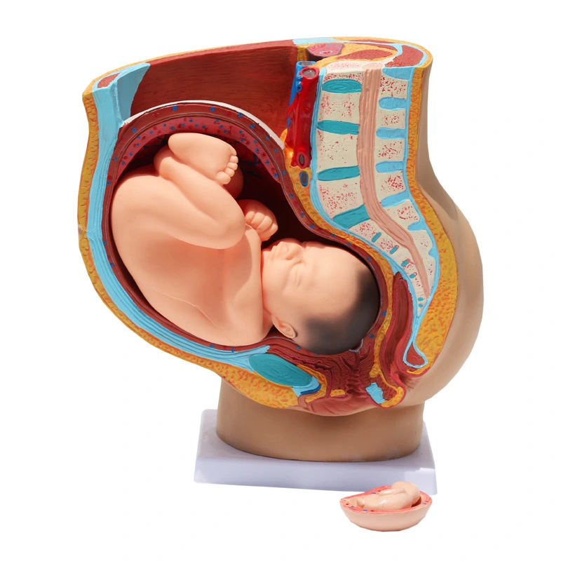
    This model is ideal for use in medical education, nursing programs, obstetrics and gynecology training, and patient education. Its realistic representation of the fetal position in the uterus and its clear depiction of the female pelvic anatomy provide valuable insight into the processes of pregnancy and birth.

    Size: life size

    Material: PVC

    3 (292)
    5 (231)

    Features

    • Realistic Representation: The model offers a detailed and realistic depiction of the pregnant pelvis and a mature fetus in the third trimester.
    • 2-Part Design: The model is split into two parts, allowing clear observation of the female pelvic anatomy and the fetal development within the uterus.

    • Detailed Pelvic Anatomy: The pelvic region, including the uterus, cervix, pelvic bones, and birth canal, is meticulously reproduced for accurate educational representation.

    Functions

    Pregnancy Pelvis with Mature Fetus - 2 Parts model serves several important functions in medical education and patient care:

    • Educational Tool: Used to teach medical students, nursing students, and obstetricians about the anatomy of the pregnant pelvis, including uterine development, fetal growth, and changes that occur during pregnancy.
    • Fetal Positioning: The model provides a clear demonstration of the position of the fetus in the uterus during the third trimester, helping students and professionals understand how the fetus is situated before birth.
    4 (247)
    2 (298)

    Benefits

    • In-Depth Study of Pregnancy: Provides an in-depth, hands-on way to learn about pregnancy, fetal development, and pelvic changes.
    • Clear Visualization: Offers a clear and accurate visual representation of the female reproductive system during pregnancy, making complex concepts easier to understand.

    • Detailed Fetal Representation: The mature fetus allows students and healthcare professionals to understand the positioning and growth of the fetus near the time of delivery.

    Applications

    • Medical Education: Ideal for obstetrics, gynecology, and nursing programs to teach students about pregnancy and female pelvic anatomy.
    • Obstetric Training: Used by obstetricians to demonstrate fetal positioning and the anatomy of the birth canal for labor and delivery training.

    • Patient Education: Can be used in prenatal classes or during patient consultations to explain the changes occurring in the female body during late-stage pregnancy.
    1 (290)

    FAQ

    1
    What parts are included in the model?
    The model consists of two parts: the pregnant pelvis with uterus and cervix, and the mature fetus.
    2
    Is this model suitable for teaching fetal positioning?
    Yes, the model is an excellent resource for teaching fetal positioning and understanding the fetus’s growth and position in the uterus.
    3
    Can this model be used for patient education?
    Yes, it is a great tool for patient education, particularly in prenatal and obstetrics settings.
    4
    Does the model provide a detailed view of the birth canal?
    Yes, it accurately represents the birth canal, including the cervix and pelvic bones, showing how the fetus is positioned in the late stages of pregnancy.
    5
    Can the model be used to demonstrate labor and delivery?
    Yes, it is useful for demonstrating how the fetus moves through the birth canal during delivery.
    6
    Can this model be used in obstetrics practice?
    Yes, it is perfect for obstetricians to demonstrate the anatomy of pregnancy and the process of labor.
    Get in touch with us
    just leave your email or phone number in the contact form so we can send you a free quote for our wide range of designs
    Related Products
    no data

    We provide targeted, flexible ,one-stop medical equipment complete solutions tailored for hospitals, laboratories, clinics and etc.

    Contact Us

    Attn: Alisa Wang 

    Mob/Whatsapp/Wechat: +86 17817884386

    Email: alisa@innocommed.com

    Add: Rm310, Building B, Fangdajing Creative Community, No.88 Guangshan San Road, Guangdong,China.

    Copyright © 2025 Guangzhou INNORKOM Medical Limited | Sitemap  | privacy policy
    Contact us
    whatsapp
    contact customer service
    Contact us
    whatsapp
    cancel
    Customer service
    detect