


Natural Uterus Model | INNORKOM
Natural Uterus Model is a detailed, anatomical representation of the human uterus, designed to provide a clear and accurate visual understanding of the organ's structure, function, and role in reproductive health. This model is commonly used in educational, medical, and clinical settings to demonstrate the anatomy and physiological processes associated with the uterus, including menstruation, pregnancy, and childbirth. It serves as an essential tool for students, healthcare professionals, and patients alike, helping to enhance the understanding of the female reproductive system.
5.0
Brand Name:
INNORKOM
Model Number:
INC-EUM10
Place of Origin:
China
Payments:
L/C, D/A, D/P, T/T, Western Union, Money Gram, OA
Lead Time:
1-1(pieces):9(days),>1(pieces):To be negotiated(days)
shipping:
Support Express/Air freight/Sea freight
design customization
Please fill out the form below to request a quote or to request more information about us. Please be sure to upload customized requirement documents or pictures, and we will get back to you as soon as possible with a response. we're ready to start working on your new project, contact us now to get started.
Please fill out the form below to request a quote or to request more information about us. please be as detailed as possible in your message, and we will get back to you as soon as possible with a response. we're ready to start working on your new project, contact us now to get started.
Product Introduction
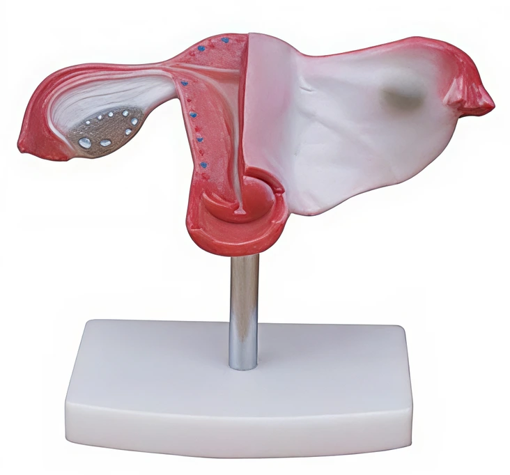
With the coronal section this model shows:
1) Uterus
2) The cervix is divided into a supra-vaginal portion and a vaginal portion
3) The ligaments of the uterus are eight in number
4) The fallopian tubes
5) The ovaries
Features
- Accurate Anatomical Representation: The model offers a highly detailed, life-like representation of the uterus, including the endometrium (lining of the uterus), myometrium (muscular layer), and serosa (outer layer).
- Removable Parts: Many models include detachable or removable sections, such as the uterus or ovaries, to allow for more in-depth examination of the organ's internal structure and its relationship with other reproductive organs.
- Color-Coded Components: Different parts of the uterus are often color-coded to make learning easier, such as highlighting the blood vessels, muscles, and connective tissues.
Function
- Menstruation: The model can illustrate how the endometrium thickens and sheds during the menstrual cycle.
- Pregnancy: It can show how the uterus accommodates a developing fetus and the changes it undergoes during pregnancy and childbirth.
- Fetal Development: By using the model, healthcare professionals can explain how the uterus supports the growth and development of a fetus.
- Anatomical Study: It serves as a valuable tool for understanding the anatomy of the uterus, including its position, surrounding organs, and the vascular system.
Benefits
- Educational Value: The model offers an interactive and visual learning experience, making it an ideal tool for teaching anatomy, reproductive health, and gynecology.
- Medical Training: It is commonly used in medical schools and clinics to educate students and healthcare professionals about the female reproductive system, especially during courses on obstetrics, gynecology, and fertility.
- Patient Education: Doctors and nurses can use the model to explain uterine conditions (such as fibroids, endometriosis, or uterine cancer) and surgical procedures like hysterectomies or cesarean sections to patients in a clear and understandable manner.
Applications
- Medical Education: Used in anatomy, gynecology, and obstetrics courses to help students and medical professionals understand uterine structure and function.
- Patient Counseling: Healthcare providers use the model to explain medical conditions related to the uterus (like fibroids, adenomyosis, or prolapse) and discuss possible treatment options.
- Surgical Training: Surgeons and medical trainees use the model to familiarize themselves with the uterus before performing operations such as hysterectomies or laparoscopies.
FAQ
1
What organs are included in the Natural Uterus Model?
The model primarily focuses on the uterus, but may also include surrounding structures such as the ovaries, fallopian tubes, cervix, and bladder.
2
What materials are used in the model?
The model is usually made from durable, life-like materials such as silicone, rubber, or plastic, which replicate the texture of human tissue.
3
Can the parts of the model be removed?
Many models offer removable or detachable parts, such as the uterus or ovaries, to allow for a more detailed exploration.
4
Is the model life-sized?
The model is often available in both life-sized and scaled-down versions for different educational needs.
5
How is the Natural Uterus Model useful in medical education?
It helps students and healthcare professionals understand the anatomy of the uterus, its function in reproduction, and various conditions affecting the uterus.
{{item.score}} Stars
{{item.pre}}%
{{item.nickname ? (item.nickname.slice(0, 2) + '*****') : item.source === 1 ? 'mall buyer' : '--'}}
{{item.comment_time}}
Review in the {{item.country}}
{{itemAttr.params_key}}: {{itemAttr.params_value}}
Get in touch with us
just leave your email or phone number in the contact form so we can send you a free quote for our wide range of designs
We provide targeted, flexible ,one-stop medical equipment complete solutions tailored for hospitals, laboratories, clinics and etc.
Useful Links
Contact Us
Attn: Alisa Wang
Mob/Whatsapp/Wechat: +86 17817884386
Email: alisa@innocommed.com
Add: Rm310, Building B, Fangdajing Creative Community, No.88 Guangshan San Road, Guangdong,China.