


Magnified Uterus Model | INNORKOM
Magnified Uterus Model is an enlarged, highly detailed representation of the female uterus designed for educational and clinical purposes. This model magnifies the structure of the uterus to offer a more comprehensive view of its internal anatomy, enabling users to explore its detailed features, such as the layers of the uterine wall, the cervix, and the blood vessels. It is a valuable tool for understanding the complex functions of the uterus in reproduction, menstruation, and pregnancy.
5.0
Brand Name:
INNORKOM
Model Number:
INC-EUM11
Place of Origin:
China
Payments:
L/C, D/A, D/P, T/T, Western Union, Money Gram, OA
Lead Time:
1-1(pieces):9(days),>1(pieces):To be negotiated(days)
shipping:
Support Express/Air freight/Sea freight
design customization
Please fill out the form below to request a quote or to request more information about us. Please be sure to upload customized requirement documents or pictures, and we will get back to you as soon as possible with a response. we're ready to start working on your new project, contact us now to get started.
Please fill out the form below to request a quote or to request more information about us. please be as detailed as possible in your message, and we will get back to you as soon as possible with a response. we're ready to start working on your new project, contact us now to get started.
Product Introduction
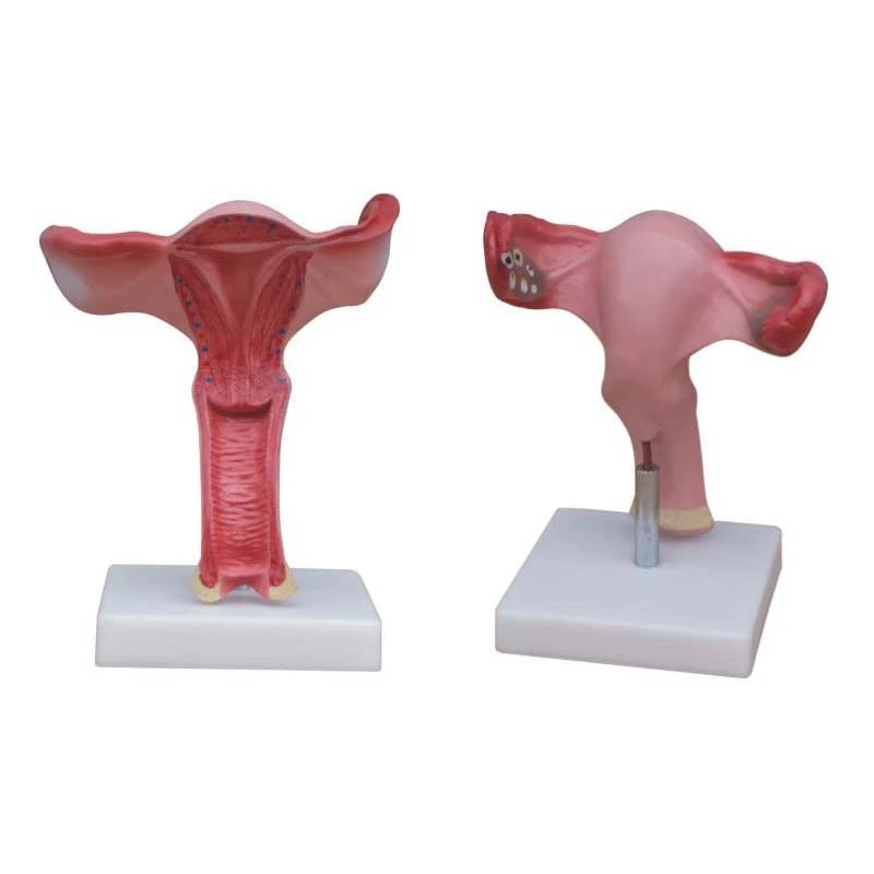
With the coronal section this model shows:
1) Uterus
2) The cervix is divided into a supra-vaginal portion and a vaginal portion
3) The ligaments of the uterus are eight in number
4) The fallopian tubes
5) The ovaries
Product Display
6 (148)
7 (82)
8 (65)
Product Advantages
Enhanced Learning Experience
The magnified scale of the model allows for a more thorough understanding of uterine anatomy, offering a clear visual representation of complex structures that are often difficult to comprehend in standard-sized models.
Improved Patient Education
Healthcare providers use the model to explain uterine conditions, surgical procedures, or reproductive health in a clear, accessible way to patients, helping them better understand their diagnoses and treatment options.
Visualizing Uterine Pathologies
The model makes it easier to identify and demonstrate common uterine disorders, such as fibroids, polyps, or abnormal growths, which can impact fertility and menstrual health.
Product Detail
- Enlarged Scale: The model is magnified to provide a closer, more detailed view of the uterus and its internal structures, allowing for an in-depth examination of anatomical details that are difficult to see in a normal-sized model.
- Detailed Uterine Layers: The magnified model highlights the three key layers of the uterus: the outer serosa, the middle myometrium (muscle layer), and the inner endometrium (lining).
- Visible Blood Vessels: The model often features color-coded blood vessels, such as the uterine artery and veins, to demonstrate the rich blood supply to the uterus and its role in reproductive health.
Material Introduction
- Anatomical Education: It provides a clear and detailed view of the uterus, aiding in the understanding of its anatomical structure, layers, and relationship with surrounding organs.
- Reproductive Health: The model helps explain the role of the uterus in menstruation, conception, and pregnancy. It shows how the uterine lining changes during the menstrual cycle and supports fetal development during pregnancy.
- Pathological Study: The magnified model is useful for identifying and studying common uterine pathologies, such as fibroids, adenomyosis, or endometriosis, and their impact on the function of the uterus.
FAQ
1
What is the purpose of a magnified uterus model?
The model is designed to offer a detailed, enlarged view of the uterus to help individuals better understand its structure, function, and potential health issues.
2
How is the magnified uterus model different from a regular model?
The magnified version provides an up-close, detailed look at the internal structures of the uterus, such as the layers of the uterine wall and blood vessels, which are often difficult to see in standard models.
3
What are the main features of the magnified uterus model?
Key features include detailed anatomical layers (serosa, myometrium, and endometrium), visible blood vessels, and often removable parts to view internal structures like the cervix or ovaries.
4
Can the model be used to explain uterine conditions?
Yes, the magnified uterus model can show conditions such as fibroids, endometriosis, and adenomyosis, helping patients and healthcare providers understand these conditions in greater detail.
5
Can the magnified uterus model assist in understanding pregnancy?
Yes, the model can demonstrate how the uterus accommodates a developing fetus and the changes it undergoes during pregnancy.
{{item.score}} Stars
{{item.pre}}%
{{item.nickname ? (item.nickname.slice(0, 2) + '*****') : item.source === 1 ? 'mall buyer' : '--'}}
{{item.comment_time}}
Review in the {{item.country}}
{{itemAttr.params_key}}: {{itemAttr.params_value}}
Get in touch with us
just leave your email or phone number in the contact form so we can send you a free quote for our wide range of designs
We provide targeted, flexible ,one-stop medical equipment complete solutions tailored for hospitals, laboratories, clinics and etc.
Useful Links
Contact Us
Attn: Alisa Wang
Mob/Whatsapp/Wechat: +86 17817884386
Email: alisa@innocommed.com
Add: Rm310, Building B, Fangdajing Creative Community, No.88 Guangshan San Road, Guangdong,China.